
The upper part of the ankle joint comes from the tibia (shin) and the fibula (outer leg). The tibia forms the front, rear and inner part of the ankle joint. The lower fibula forms the outer part of the ankle joint. The ends of these bones are called malleoli. There are two malleoli on the tibia (medial and posterior) and one on the fibula (lateral).
Ankle fractures occur when the malleoli are broken. These fractures are very common. Ankle fractures can happen after falls, car accidents or twisting of the ankle. One, two or all three malleoli can be broken.
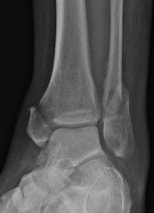
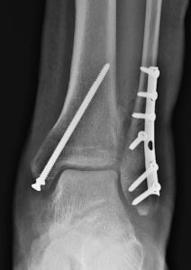
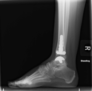
Symptoms of an ankle fracture are pain, swelling, bruising and problems with ankle motion. X-rays help show which need treatment from an orthopedic surgeon. The main goal of surgery is to get the ankle joint to heal with a normal shape. Once the ankle is put back together, the next step is to regain normal movement.
The long-term goal of repairing a broken ankle is to decrease the chances of ankle arthritis in the future. Most ankle fracture surgery involves open reduction and internal fixation (ORIF). An incision is made over the ankle to see the fractured bones. Like a jigsaw puzzle, the pieces of the broken bones are placed back together (open reduction). The broken bones are then held together (internal fixation) in this correct position with metal plates and/or screws. This internal fixation provides stability so movement can begin shortly after surgery as the ankle fracture heals.